Oshane O. Thomas, PhD

AI & Bioinformatics Scientist — Machine Learning for Medical Imaging & Morphology
Seattle, WA • On-site/Remote/Hybrid • GmailLinkedInGitHub

View the Project on GitHub

Skills banner

📚 Publications

Peer-Reviewed Publications

Trustworthy detection of exencephaly in high-throughput micro-CT embryo screens with focal-loss transformers

Thomas, O. O., Roston, R., Shen, H., & Maga, A. M. (2025). BioRxiv (In Review: PLOS Computational Biology). https://doi.org/10.1101/2025.08.12.669840

SlicerMorph photogrammetry: An open-source photogrammetry workflow for reconstructing 3D models

Thomas, O. O., Zhang, C., & Maga, A. M. (2025). Biology Open, 14, bio062126. https://doi.org/10.1242/bio.062126

Leveraging descriptor learning and functional map-based shape matching for automated anatomical landmarking in mouse mandibles

Thomas, O. O., & Maga, A. M. (2025). Journal of Anatomy, 00, 1–17. https://doi.org/10.1111/joa.14196

Automated morphological phenotyping using learned shape descriptors and functional maps: A novel approach to geometric morphometrics

Thomas, O. O., Shen, H., Raaum, R. L., Harcourt-Smith, W. H. E., Polk, J. D., & Hasegawa-Johnson, M. (2023). PLOS Computational Biology, 19(1), e1009061. https://doi.org/10.1371/journal.pcbi.1009061

💼 Experience

Postdoctoral Fellow

Seattle Children’s Research Institute & The Imageomics Institute, Seattle, WA (09/2023–Present)

  • Built end-to-end pipelines for micro-CT embryo analysis (NIfTI), covering segmentation, classification, detection, and registration; packaged with Docker/MLflow for reproducibility and cloud training.
  • Designed and published a 3D Slicer extension integrating SAM-based segmentation with WebODM photogrammetry, cutting 3D reconstruction error by ~15% on fragile specimens; enabled volume-to-mesh workflows for downstream analysis.
  • Developed focal-loss Vision Transformer models for neural tube defect (exencephaly) detection under extreme class imbalance; improved AUPRC and stabilized XAI saliency.
  • Automated functional-map-based landmarking for 3D morphology, boosting reproducibility and throughput; integrated experiment tracking (MLflow) and artifact versioning.
  • Led monthly training on medical imaging ML (3D CNNs, ViTs, SSL, Photogrammetry), supporting cross-functional teams.

Research Assistant

Speech Technology Group, Coordinated Science Lab, Grainger College of Engineering, UIUC (09/2021–08/2023)

  • Adapted Transformer architectures with XAI for high-dimensional gait/kinematics; delivered interpretable, production-grade models and validation reports.

Research Assistant

Evolutionary Biomechanics Lab, Department of Anthropology, UIUC (09/2017–12/2018)

  • Automated 3D trait extraction via geometry processing on surface scans; mentored students in Python computer vision workflows.

Research Assistant

Immunology and Genomics Lab, Department of Anthropology, UIUC (09/2016–05/2017)

  • Quantified skeletal traits from micro-CT; authored an R package linking genotype–phenotype at scale.

🚀 Featured Work

Trustworthy Exencephaly Detection with Focal‑Loss Transformers

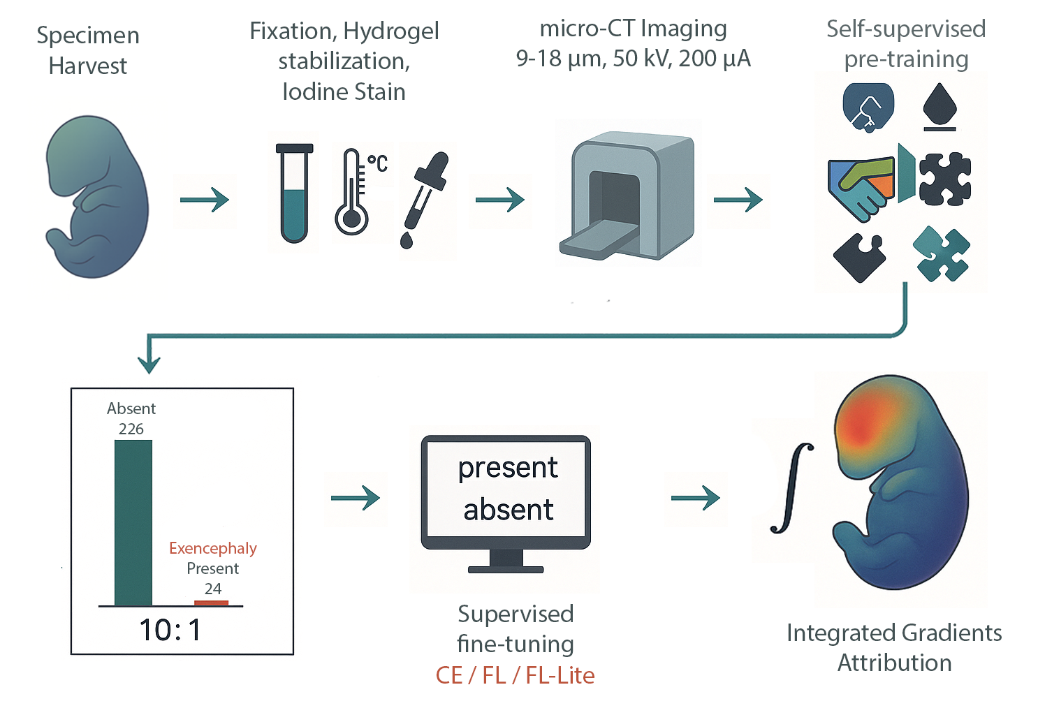
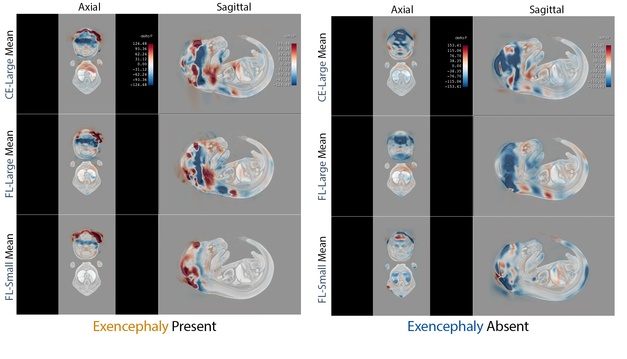
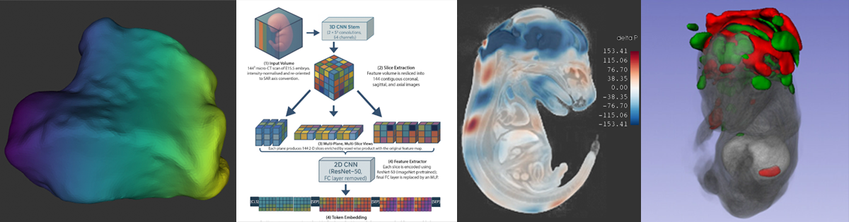
IMPC/KOMP‑style whole‑embryo iodine‑contrast micro‑CT (diceCT) screens search for lethal malformations across knockout lines—but positives are rare (~10%), a recipe for brittle classifiers and noisy saliency. We built an imbalance‑aware transformer that pairs focal loss with capacity control and seed ensembling, and we evaluated explanation quality—not just ROC. On 253 E15.5 volumes (24 exencephaly), all 15 models achieved 0.996 ± 0.002 accuracy; focal loss cut saliency entropy by up to 1.5 bits and doubled cross‑seed Dice, focusing attribution on the malformed cranial vault.

This urns attribution/saliency from impressionistic heatmaps into anatomy‑specific, reproducible evidence—without voxel labels—using only atlas registration and standard preprocessing.

Our approach delivers trustworthy, high‑throughput phenotyping for large mouse genetics programs, triaging defects with near‑perfect sensitivity and reproducible explanations. The recipe generalizes to other malformations and cohorts, offering a practical template for interpretable 3D classification under severe class imbalance.

Decoding Shape Diversity: An Autoencoder-Based Morphospace for Comprehensive Biological Shape Analysis

We build an interactive morphospace for whole‑surface anatomy. Using morphVQ functional‑map correspondences (landmark‑free, no shared mesh), we train a DISCO‑AE mesh autoencoder to learn a smooth latent space that decodes to valid surfaces. Moving along an axis warps a template and paints local deformation fields, showing which regions expand, contract, or bend—and why groups differ.

Unlike PCA on sparse landmarks, this covers every vertex, is stable across mesh resolutions, and yields anatomy‑faithful, continuous trajectories with PCA‑like interpretability.

This enables cohort‑scale, interpretable exploration for comparative anatomy, preclinical phenotyping, and device design; supports QC, hypothesis generation, and downstream analysis (exportable meshes/fields). Built to plug into SlicerMorph for reproducible, large‑dataset workflows.

DISCO-AE Citation: S. Hahner, S. Attaiki, J. Garcke and M. Ovsjanikov, “Unsupervised Representation Learning for Diverse Deformable Shape Collections,” 2024 International Conference on 3D Vision (3DV), Davos, Switzerland, 2024, pp. 1594–1604, doi: 10.1109/3DV62453.2024.00158.

SlicerMorph Photogrammetry: SAM‑NodeODM Pipeline for High‑Fidelity 3D Specimens

This project delivers a fully open, end‑to‑end photogrammetry workflow inside 3D Slicer that integrates automatic image masking via the Segment Anything Model (SAM) with surface reconstruction using OpenDroneMap’s NodeODM, wrapped in a user‑friendly Slicer extension. The module unifies image import, batch/single‑image masking, optional scaling via ArUco/GCPs, reconstruction, and direct model import for morphometric analysis.

Goals were twofold: streamline background removal and improve geometric accuracy, especially around delicate cranial structures. Using 14 mountain‑beaver skulls with micro‑CT as reference, the updated workflow lowered mean surface distance and RMSE by ~10–15% across specimens versus our prior open‑source pipeline, while Hausdorff changed little—evidence that gains were global rather than driven by a few outliers. Qualitatively, thin elements (e.g., zygomatic arches, orbital rims) showed fewer breaks, clogs, and smoothing artifacts. A Taguchi L16 design efficiently explored NodeODM parameters (e.g., mesh‑octree‑depth, mesh‑size, feature/pc quality), yielding a reproducible configuration that balances fidelity and runtime; all job configs are saved as JSON for auditability.

Findings: (1) Automated SAM masking markedly reduces manual cleanup and operator bias; (2) tuned NodeODM reconstruction produces smoother, more anatomically faithful meshes; and (3) the one‑ecosystem approach (mask → reconstruct → analyze) accelerates research hand‑offs to SlicerMorph morphometrics. Broader impact spans natural‑history collections, ecology/evolution studies, teaching/outreach, and large‑cohort digitization where cost, licensing, and throughput matter. Cloud‑ready deployment (MorphoCloud) supports GPU‑accelerated processing without local DevOps overhead.

Spatially & Spectrally Consistent morphVQ: Functional‑Map Landmarking for Mouse Mandibles

Manual landmarking slows morphometrics and bakes in bias. This paper delivers an automated, unsupervised pipeline that learns dense correspondences across mandibles and then reads off canonical landmarks, eliminating rigid pre‑alignment.

Using orientation‑preserving complex functional maps, DiffusionNet descriptors, and spatial/spectral cycle‑consistency, the model places landmarks with accuracy competitive with MALPACA while running faster and robust on small training sets. Validation spans 425 hemi‑mandibles from micro‑CT.

Landmarks encode a hypothesis; changing that hypothesis usually means re‑annotating everything. Here, once correspondences are learned, you can regenerate any landmark set—classical or custom—without relabeling, turning landmarking into a query rather than a bottleneck. This enables scalable morphometrics for evo‑devo genetics, QTL mapping, and phenome‑wide screens, and translates to dental/orthopedic planning and preclinical pipelines where fast, consistent, anatomy‑aware points are essential.

morphVQ: Functional-Map Morphometrics for Automated Phenotyping

Most 3D shape analysis still relies on experts clicking landmarks—slow, biased, and blind to much of the surface. morphVQ replaces that with an automated pipeline that learns dense correspondences across whole meshes and summarizes shape as surface‑wide expansions, contractions, and angle changes—compact, interpretable descriptors.

It scales unbiased quantification to large cohorts. On primate bones, morphVQ matched landmark pipelines while capturing more detail and running faster. It also projects “where the differences are” back onto the surface for review.

Beyond comparative anatomy, teams in medical imaging, preclinical phenotyping, surgical planning, and device design can trade manual clicks for reproducible, full‑surface metrics. For ML teams, it showcases geometric deep learning for correspondence learning and interpretable features.

🛠️ Skills and Expertise

Tech stack at a glance:

  • Python
  • PyTorch
  • TensorFlow/Keras
  • Docker
  • AWS
  • GCP
  • SQL
  • NIfTI/DICOM

Modalities & Data Ops

  • Formats & ecosystems: DICOM, NIfTI; whole‑embryo diceCT; surface meshes (OBJ/PLY).
  • Pipelines: end‑to‑end data loaders, artifact/version tracking, automated reports for accuracy & calibration.

CV/ML Tasks/Algorithms & XAI

  • Core tasks: 3D segmentation, classification, anomaly detection, and automatic landmarking.
  • Architectures: Vision Transformers, SwinUNETR, 3D U‑Net/UNet++, geometric deep learning (functional maps, DiffusionNet), mesh autoencoders.
  • Techniques: Integrated Gradients, Layerwise Relevance Propagation.

Tooling & Platforms

  • Med-imaging & 3D: 3D Slicer/SlicerMorph, VTK/ITK, OpenCV, trimesh.
  • Photogrammetry: OpenDroneMap (NodeODM).
  • Programming: Python, R/Rcpp, C/C++, SQL; PyTorch, PyTorch-Geometric, TensorFlow/Keras; scikit-learn, pandas, NumPy, statsmodels.

MLOps & Reproducibility

  • Stack: Docker, MLflow, WandB, Git/GitHub Actions; AWS/Azure/GCP for GPU training & inference.
  • Practices: experiment tracking, artifact versioning, config-as-code, CI for data/metrics drift, model cards.

Open‑Source & Extensions

  • SlicerPhotogrammetry (SAM→NodeODM end‑to‑end), MorphoSourceImport, SkyscanReconImport, HiResScreenCapture, SlicerScriptEditor — modules adopted for faster, reproducible 3D data processing and visualization.

Stats & Modeling

  • Bayesian & classical inference; variational methods; predictive modeling; statistical shape analysis; evaluation beyond accuracy (AUPRC, calibration, uncertainty).

📄 Curriculum Vitae

Download Full CV (PDF)